
Fetal Heart Scan

Known medically as Early Fetal Echocardiography, or simply Early Echo, this specialised scan offers a detailed view of your baby’s heart as early as 12 weeks into pregnancy.

We highly recommend this scan for all babies with increased nuclal translucency (NT) measurements, fetal anomalies, or other unusual findings detected at 11-13 weeks scan. This early scan is crucial for assessing your baby’s heart health, providing peace of mind, and allowing for early intervention if needed.

Expectant mother points at a fetal heart development chart after getting the Early Baby Heart Scan, also know as Early Fetal Echocardiography, done.

Early Reassurance Of Your Baby’s Heart

Heart defects (cardiac anomalies or congenital heart disease/CHD) are the most common human anomalies. They are even more common than Down’s syndrome and can sometimes be critical for your baby. 

This is why Dr Fred Ushakov recommends our Early Fetal Heart Scan as this test is specifically designed to examine your baby’s heart. It is a vital tool for the early detection of CHD, ensuring early intervention and the best possible care for your little one’s heart.

Our Early Fetal Echocardiography goes beyond heart assessment and is a second opinion pregnancy scan, offering a thorough examination of your baby’s overall health from head to toe. This comprehensive approach is crucial for detecting any anomalies and provides vital health information, especially regarding heart conditions. 

Baby with increased NT (6.0mm vs 3.5mm cutoff) and severe heart defect diagnosed by our Early Baby Heart Scan, or Early Fetal Echocardiography, at 12 weeks

Baby with increased NT (6.0mm vs 3.5mm cutoff) and severe heart defect diagnosed by Early Fetal Echocardiography at 12 weeks

Who will benefit from Early ECHO?

There are well-recognised maternal and fetal conditions strongly associated with increased risk for the baby to have a cardiac anomaly. If you, your family, or your baby have any of those risk factors you may wish to perform Early Fetal Echocardiography (which also includes Early Fetal Scan).

Common risk factors of congenital heart defects/CHD:

  • Baby’s increased nuchal translucency (NT) of 3.5 mm or more

  • Suspected heart abnormality at 11-13 weeks scan

  • Abnormal NIPT or invasive test (CVS) results

  • Ultrasound findings at 11-13 weeks scan like fetal anomalies, collections of fluid in the body, single umbilical artery, and others

  • Abnormal baby’s heartbeat (irregular, too fast, or too slow)

  • Tricuspid regurgitation (TR) or abnormal flow in ductus venosus (DV) for your baby

  • If you have diabetes (especially before being pregnant)

  • If your baby was conceived by in vitro fertilization (IVF), including intracytoplasmic sperm injection (ICSI)

  • Family history of heart anomalies

  • If you were taking medicines or other substances known to increase the risk for baby’s heart defects

Be sure about the heart from 12 weeks

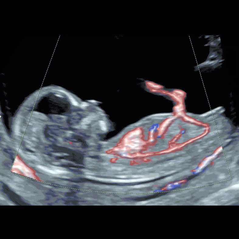
At 12-13 weeks, the fetal heart is about the size of corn grain, and with our sophisticated ultrasound and Colour Doppler technology, we meticulously examine the fetal heart. We assess the heart’s and stomach’s position, the size and presence of all four chambers, and the two great arteries. The advanced Colour Doppler technique, a pivotal component of our Early Fetal Heart Scan, allows us to meticulously assess the blood flow within your baby’s heart and across the cardiac valves.

A recent study from Oxford suggested that up to 80% of prenatally detectable heart anomalies can be diagnosed in high-risk populations in the 1st trimester (Karim, et al, 2022). Normal results of our 12-18 weeks Fetal Echocardiography will provide the earliest reassurance that your baby’s heart develops normally.

Our Early Fetal Echocardiography offers a unique advantage by not only evaluating the fetal heart but also conducting an advanced and thorough structural examination of the baby’s overall health. Through this comprehensive assessment, we meticulously examine all the parts and organs of the baby, aiming to detect any associated anomalies. This advanced diagnostic capability is invaluable as it provides crucial information to parents about their baby’s health, particularly concerning the condition of the heart.

Baby's heart, main arterial and venous vessels and umbilical cord blood flow by our special colour Doppler technology. Image done during London Pregnancy Clinic's Early Baby Heart Scan, or Early Fetal Echocardiography, at 12 weeks

Baby’s heart, main arterial and venous vessels and umbilical cord blood flow by our special colour Doppler technology at 12 weeks

Frequently Asked Questions

You have a question? We have an answer.

Who performs the Fetal Heart Scan in London Pregnancy Clinic?

All Fetal Echocardiography Scans (Fetal Heart Scan) are performed by Dr Fred Ushakov, who is an internationally recognised expert and teacher in this field. Dr Fred’s mission is to empower ultrasound practitioners with ‘proper education in fetal echocardiography,’ aiming to boost the detection rate of CHD and thus ‘save many lives.’

He has organised, chaired and spoken at multiple conferences with London School of Ultrasound on the subject of Fetal Echo during the Annual EFScan Conferences.

What is the difference between Fetal Heart Scan and Fetal Echocardiography

At the London Pregnancy Clinic, “Fetal Heart Scan” and “Fetal Echocardiography” refer to the same procedure. The term “Fetal Heart Scan” is used to help expectant mothers understand that the scan is specifically focused on the heart, emphasising its specialised nature in assessing the baby’s heart health early in the pregnancy. This naming convention aims to make the purpose of the scan clearer and more relatable for mothers-to-be.

What is procedure of Early Fetal Echocardiography looks like?

For you, fetal echocardiography will resemble a regular ultrasound scan. You will see your baby’s beating heart on our special screen and we will explain to you what structures we check.

In the majority of cases, we need to use both transabdominal and transvaginal approaches to get the best quality images of your baby’s heart. Understandably you can choose to avoid a transvaginal scan, however, in some cases the information regarding the baby’s heart will be incomplete or inaccurate without this examination.

Are you scanning also other baby’s organs or just the heart?

We believe that the heart together with the brain is the most important part of the humans. Early Fetal Echocardiography has special focus on fetal heart, however we perform comprehensive top-to-toe examination of all structures for your baby. 

Can I have baby’s heart images and clips from the scan?

Yes, of course. We use Tricefy which is a secure cloud system to upload images and clips from your baby’s heart scan to be viewed by your doctor. 

Tricefy also allows you to download the images and clips of the heart and share them with your fetal medicine or obstetric consultant or fetal cardiologist. By your special request we can also directly share securely those images and clips with your doctor. For this you will need to provide his/her secure email address.

You will also receive a hard and soft copy of your detailed ultrasound and early fetal echocardiography report as well as some high-quality colour printouts of the baby for you to take home with you.

Are we helping in fetal Echo training of other professionals?

Yes, definitely. The website of the London School of Ultrasound associated with our clinic has its unique name fetalechocardiography.com. Not only are we internationally recognised experts in performing Early Fetal Echocardiography, but we also have a long-standing record of training this technique to professionals in the UK and around the world.

Can you diagnose all fetal heart anomalies at 1st trimester?

In the first trimester we can diagnose about 80% of severe heart defect which can be recognised prenatally (before birth).

We cannot diagnose isolated ventricular heart defects, progressive abnormal development of the valves and some other heart anomalies.

I have nuchal translucency (NT) measurement 3.0 mm. Do I need to have Fetal Echo?

Generally yes, however it is still does not considered an indication for Fetal Echocardiography in UK.

American Institute of Ultrasound in Medicine (AIUM) in its Practice Parameter for the Performance of Fetal Echocardiography considers ‘greater-than-normal nuchal translucency measurement between 3.0 and 3.4 mm’ as an indication for Fetal Echo.

What are the most common indications for fetal echocardiography?
  • increased nuchal translucency (NT) for the fetus
  • inconclusive or abnormal heart scan by sonographer
  • family history of heart defects
  • maternal diabetes
  • maternal medications
What is the difference between our Early Fetal Scan and Fetal Echocardiography?

Early Fetal Echocardiography included both:

  • special comprehensive examination of the heart and great vessels (heart connections)
  • Early Fetal Scan
Can you technically perform Early Fetal Echocardiography at 12 weeks in 100% cases?

No, unfortunately sometimes fetus stays in awkward position and we cannot get all the views of the heart. In those cases we will rescan you for free later in pregnancy (usually in 2 weeks time).

I have low risk pregnancy and normal results of NIPT, can I do Early Fetal Echo?

Yes, we can perform Early Fetal Echocardiography for every mother, who is anxious regarding normality of her baby’s heart.

Unfortunately, vast majority of heart defects are diagnosed in babies from low-risk families.

How common are fetal heart anomalies in UK?

Cardiac anomalies occur very frequently. According to NHS portal congenital heart disease is one of the most common types of birth defect, affecting almost 1 in 100 babies born in the UK.

British Heart Association quotes that each day, around 13 babies in the UK are diagnosed with congenital heart disease. 


We are very healthy parents, can our baby have severe heart defect?

Unfortunately, yes. Every baby has a chance to be born with severe heart anomaly.

What will happen if my baby will have heart defect diagnosed by Early Fetal Echo?

If we will diagnose fetal cardiac anomaly for your baby you will need to be counselled by Fetal/Paediatric Cardiologist. Fetal Medicine Units in different London hospitals have various arrangements for this type of consultations. We will assist you with referral for Fetal Cardiologist.

What cardiac anomaly will be considered as severe or critical heart defect?

There are more than 300 different congenital heart abnormalities.

According to CDC: About 1 in every 4 babies born with a heart defect has a critical congenital heart defect (critical CHD, also known as critical congenital heart disease). Babies with a critical CHD need surgery or other procedures in the first year of life. 

What scientific evidence supports performing Early Fetal Echo?

There is new (2022) publication from Oxford researchers by Dr Karim and other. It is called ‘First-trimester ultrasound detection of fetal heart anomalies: systematic review and meta-analysis’.

Here is the link for this excellent scientific paper: Karim, et al, 2022

Can you diagnose ‘hole in the heart” at your 12-16 weeks Fetal Echocardiography?

Hole in the heart (ventricular septal defect or VSD) is very common anomaly. Small VSDs usually not recognised before birth. Usually this type of cardiac anomalies have good outcome (majority close spontaneously)

Early Fetal Echocardiography cannot diagnose isolated VSDs.

My partner was born with “hole in the heart”. Can you reassure us?

Yes, we can exclude up to 80% of severe or critical heart anomalies, which are detectable before birth.

Sadly I had termination of my previous pregnancy at 22 weeks due to abnormal baby’s heart. Now I am at 8 weeks and extremely anxious. How early you can reassure me?

You can book your Early Fetal Echo at 12 weeks. Please note that we will recommend you to have also transvaginal (internal) scan.

Can I have Early Fetal Cardiography together with NIPT?

Yes, you can have add-on NIPT (Harmony test or Invitae NIPS).

Please contact us regarding pricing of NIPT.

I have booked Early Fetal Echo in your clinic. Can I do also NIPT at that appointment?

Yes, you can have add-on NIPT (Harmony test or Invitae NIPS).

Please contact us regarding pricing of NIPT.

At 11 weeks scan at NHS sonographer has found something unusual for the baby. Would you recommend Early Echo?

Yes, inconclusive or abnormal result of 11-13 week scan commonly can be associated with heart anomaly.

What are the commonest heart defects?

According to British Heart Foundation

  • Aortic stenosis
  • Atrial septal defect
  • Coarctation of the aorta
  • Common arterial trunk
  • Complete and partial atrioventricular septal defect
  • Double inlet ventricle
  • Hypoplastic left heart
  • Large ventricular septal defect
  • Patent ductus arteriosus
  • Pulmonary atresia with intact ventricular septum
  • Pulmonary atresia with ventricular septal defect
  • Pulmonary stenosis
  • Supraventricular tachycardia
  • Tetralogy of Fallot
  • Transposition of the great arteries
  • Tricuspid atresia
Why Early Fetal Echocardiography is so expensive scan?

Early Fetal Echocardiography is the most expensive ultrasound scan. It is because of:

  • Need for highest level of expertise of the doctor, who performs Echo
  • Use of the most advanced and expensive ultrasound scanner and various transducers (we have 5 different types of transducers)
  • It is time consuming ultrasound scan
I have twins, can you do Early Fetal Echocardiography for my babies?

Yes, we are doing early fetal echo for twins. Please contact us to arrange your appointment.

Can fetus with Down’s syndrome also to have heart defect?

Yes, up to half of the babies with Down’s syndrome have heart defects. The most common cardiac anomaly for fetuses with trisomy 21 (T21) is atrioventricular septal defect  (AVSD) or hole in the middle of the heart.

Do I need full bladder for Early Fetal Echocardiography?

No, we definitely do not need full bladder. Full bladder interfere with quality of the scan. 

How fast I will get report of Early Fetal Echocardiography?

Usually immediately after the scan. We provide both hard copy and electronic record (PDF) formats.

However, if will find something unusual, the report can be delayed for few hours when we review again the images and video clips of your baby’s heart and will write complete report.

Can you perform counselling for severe heart defect?

No, we perform diagnostic services. Counselling for congenital heart defects performs Fetal/Paediatric Cardiologist.

I had already CVS, and waiting for Fetal Echo scheduled in few weeks. Can you check my baby’s heart earlier?

Yes, please book your appointment for Early Fetal Echocardiography as soon as possible.

Can you perform Early Fetal Echo at 10-11 weeks? I can’t wait for 12 weeks.

Yes. For very early fetal heart examination we use newest technology called “Slowflow HD’. In exceptional circumstances (like this) we can do fetal heat examination at 10-11 weeks. One of those situations is positive NIPT for Down’s syndrome (T21) when you may wish to know if the baby can have also heart defect. 

We would strongly recommend to repeat the Early Echo at 16 weeks due to understandable limitations of very early Echo.

How severe heart anomalies can be?

There is a spectrum of severity of heart defects. The most severe are unfortunately lethal. Others can be completely or partially correctable by open heart surgery.

Do I need to perform internal (transvaginal) scan to check my baby’s heart?

Yes, in majority of the cases we recommend to have both transabdominal and transvaginal scans.

My baby’s heartbeat was very fast. What is normal fetal heart rate at 11-13 weeks?

Fetal heart beat is normally very fast. Baby’s heart starts beating at around 6 weeks, when the embryo is just 2-5 mm in length; at that time baby’s pulse is 85-130 beats per minute (bpm). The spike of fetal heart rate occurs at 9-10 weeks, when baby’s heart contracts as fast as 150-190 bpm. 

There is gradual decrease in fetal heart rate between 10 and 14 weeks. Normally at 11-13 weeks fetal heart rate ranges from 145 to 175 bpm.

It was found that babies with chromosomal anomalies or heart defects can have abnormal heart beats.  For instance fetuses with trisomy 18 (Edwards syndrome) and triploidy have relatively slow pulse. Contrary to that the heartbeat of trisomy 13 (Patau’s syndrome) and Turner’s syndrome fetuses is relatively fast. Very slow fetal heart beat at 11-13 weeks (below 100 bpm) is strongly associated with serious cardiac anomalies.

Significant abnormalities of the fetal heart rate at 9-13 weeks: too fast, too slow or irregular are indications for Early Fetal Echocardiography.

What is ductus venosus. Do you check it at Early Fetal Echo?

Ductus venosus (DV) is an important vessel which allows oxygenated blood in the umbilical vein to bypass the liver and to provide oxygen to the fetal brain.

Yes, we check DV at the time of Early Fetal Echo.

Do you want to know more about Early Fetal Echo?

Early Fetal Echocardiography is a very special examination and it is difficult to find any reliable information about this ultrasound scan. Please contact us now to ask your question.